
What is a sight test?
A sight test is really just what it says… it is a test of how well, or not, a person can see, and a measurement of any corrective prescription that could be used to give someone the best vision possible. A sight test could involve additional tests such as visual fields and eye pressures but may not always detect underlying conditions requiring deeper investigation. A ‘sight test’ is just one part of a more comprehensive ‘eye examination’.
What is an eye examination?
An eye examination is a series of tests performed by an ophthalmologist, or an optometrist, assessing vision and ability to focus on and discern objects, as well as other tests and examinations pertaining to the eyes. Health care professionals often recommend that all people should have periodic and thorough eye examinations as part of routine primary care, especially since many eye diseases are asymptomatic.
Eye examinations may detect potentially treatable blinding eye diseases, ocular manifestations of systemic disease, or signs of tumours or other anomalies of the brain.
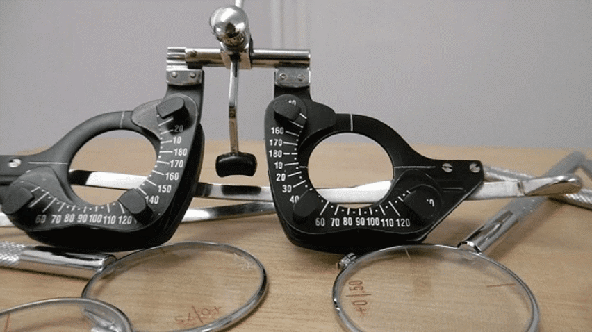
What does an eye examination consist of?
Ideally, the eye examination consists of an external examination, followed by specific tests for visual acuity, pupil function, extraocular muscle motility, visual fields, intraocular pressure and ophthalmoscopy through a dilated pupil.
A minimal eye examination consists of tests for visual acuity, pupil function, and extraocular muscle motility, as well as direct ophthalmoscopy through an undilated pupil.
What tool is used by eye examiners?
Close inspection of the anterior eye structures and ocular adnexa are often done with a slit lamp which is a table mounted microscope with a special adjustable illumination source attached. A small beam of light that can be varied in width, height, incident angle, orientation and colour, is passed over the eye.
Often, this light beam is narrowed into a vertical “slit”, during slit lamp examination. The examiner views the illuminated ocular structures, through an optical system that magnifies the image of the eye and the patient is seated while being examined, and the head stabilised by an adjustable chin rest.
This allows inspection of all the ocular media, from the cornea to vitreous, plus a magnified view of the eyelids, and other external ocular related structures. Fluorescein staining before slit lamp examination may reveal corneal abrasions or herpes simplex infection.
The binocular slit-lamp examination provides a stereoscopic magnified view of the eye structures in striking detail, enabling exact anatomical diagnoses to be made for a variety of eye conditions.
Also, ophthalmoscopy and gonioscopy examinations can also be performed through the slit lamp when combined with special lenses. These lenses include the Goldmann 3-mirror lens, gonioscopy single-mirror/ Zeiss 4-mirror lens for (ocular) anterior chamber angle structures and +90D lens, +78D lens, +66D lens & Hruby (-56D) lens, the examination of retinal structures is accomplished.
What can an eye exam find?
Nowadays, some practices also have OCT, ocular coherence tomography, which is a full 3D scan of the structures of the eye, enabling accurate and earlier diagnosis of many eye conditions as well as other conditions such as brain tumour, high blood pressure, macular degeneration, glaucoma and diabetic changes.


